
Human Tongue Anatomy Human tongue, Tongue health, Anatomy
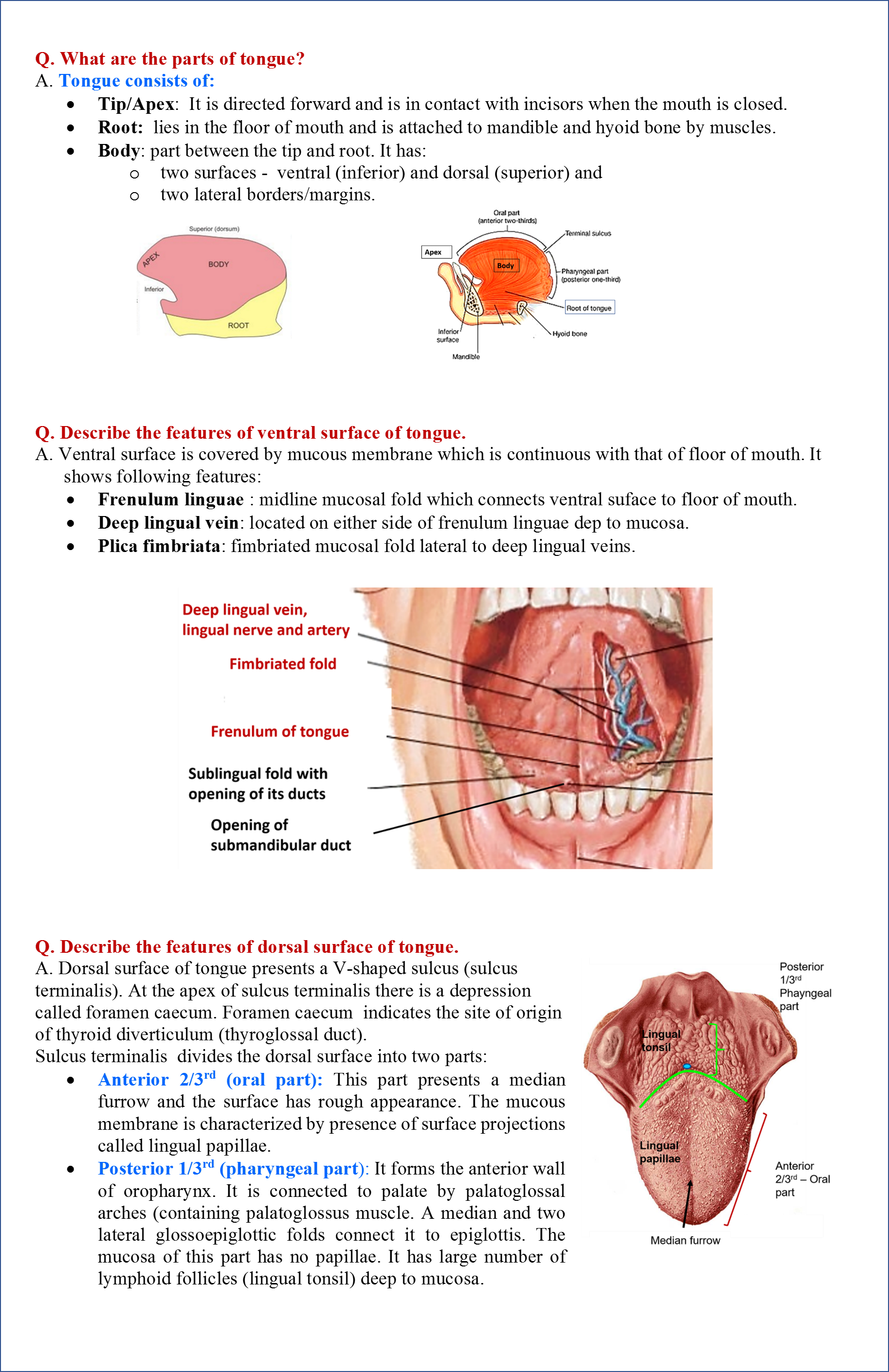
The tongue is a muscular structure as well as a sensory organ that starts developing alongside the external face around week 4 of intrauterine life.. A fully developed tongue consists of two parts, the anterior two-thirds; and posterior one-third, which is called the root of the tongue; they are separated from each other by a shallow v-shaped groove, known as the terminal sulcus.
The Tongue Anatomy Anatomical Charts & Posters
3 min read Image Source © 2014 WebMD, LLC. All rights reserved. The tongue is a muscular organ in the mouth. The tongue is covered with moist, pink tissue called mucosa. Tiny bumps called.
Tongue Anatomy Diagram Anatomical Charts & Posters
Human body Digestive System Tongue Tongue The tongue is unique in that it is the only muscle that isn't connected to bone at both ends. It is connected on one end to the hyoid bone, which is.

Cartoon of human tongue anatomy Royalty Free Vector Image
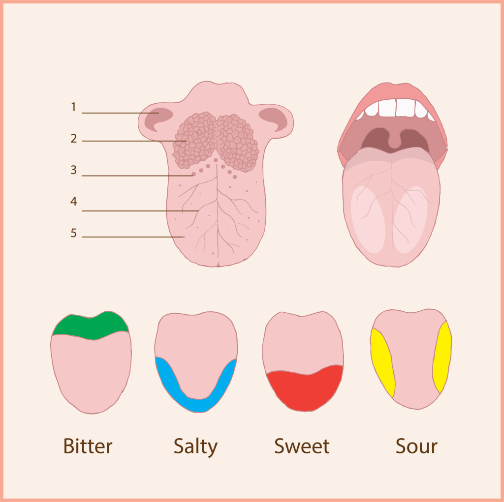
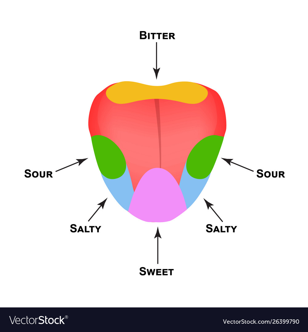
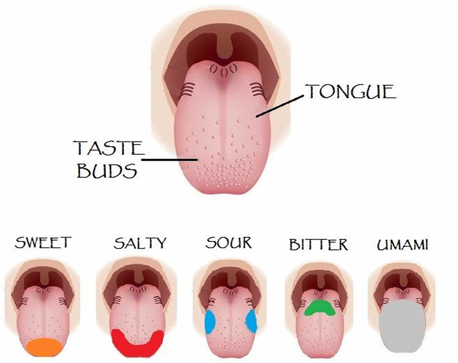
The taste buds are bulb-shaped structures responsible for taste perception, located within the lingual papillae and in the surface mucosa of the soft palate, oropharynx, epiglottis, and upper esophagus. It is the only extrinsic muscle of the tongue that is not innervated by the hypoglossal nerve but by the vagus nerve (CN X). Contraction of the.

The Tongue Diagram Quizlet
Lips and Tongue: Anatomy. The lips are the soft and movable most external parts of the oral cavity. The tongue, on the other hand, is a complex muscular structure that permits tasting and facilitates the process of mastication and communication. Together, these structures play an important role in each of these vital processes.
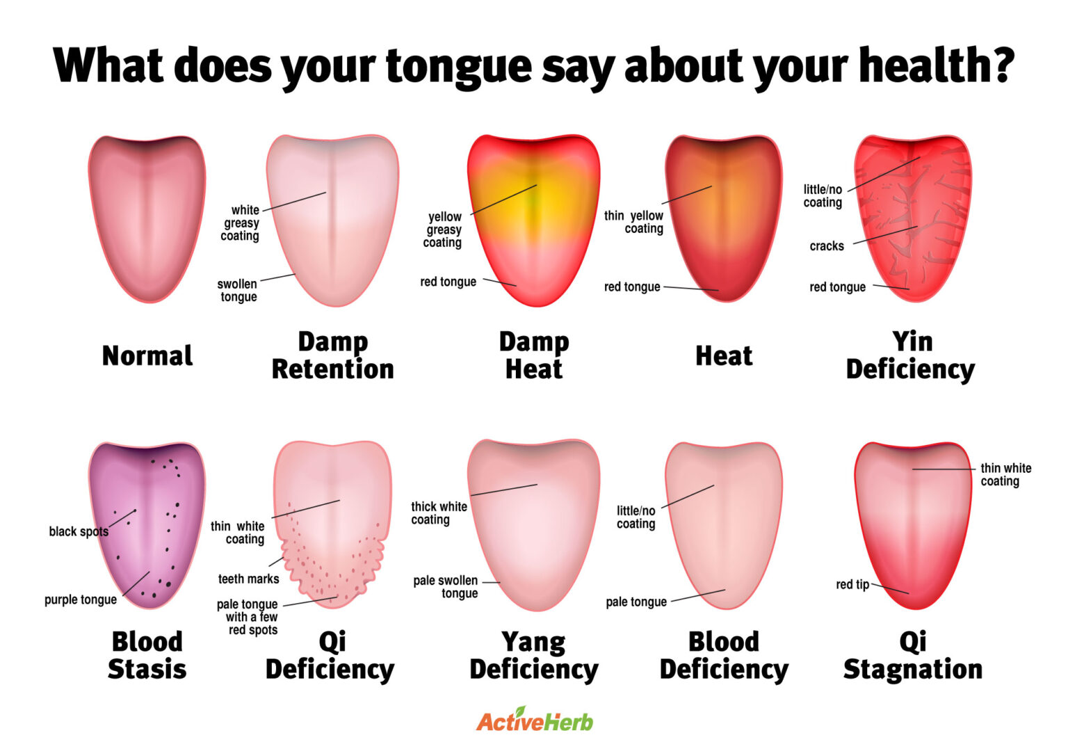
tonguediagram Activeherb Blog
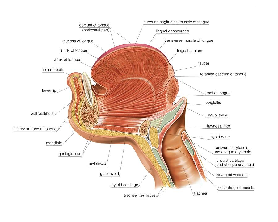
The root of the tongue is posterior and slightly vertical, forming the posterior one third of the tongue. It extends from the hyoid, epiglottis, and soft palate, to the mandible. The body forms the anterior ⅔ of the tongue, and the apex of the tongue is the most anterior end of the body. The entirety of the tongue rests on the mouth's floor.

Adult Frenectomy For Pain Relief Osteopathic ConsiderationsOsteopathy New York, P.C.
The tongue is a muscular structure in the mouth covered by mucosa whose primary functions are in mastication, taste, and speech. It can be divided into the anterior two-thirds which makes up part of the oral cavity and the posterior-third, part of the oropharynx. 1 The tongue consists of a tip, dorsal surface, ventral surface, and root.

Anatomy and Physiology Sensory Perception
Review of the tongue Embryology. The tongue begins to develop at the end of the fourth gestational week.It arises from the first, third and fourth pharyngeal arches of the pharyngeal apparatus. Initially, the first pharyngeal arch gives rise to a central tuberculum impar (also called the median lingual swelling) and the bilaterally paired lateral lingual swellings.
The Tongue
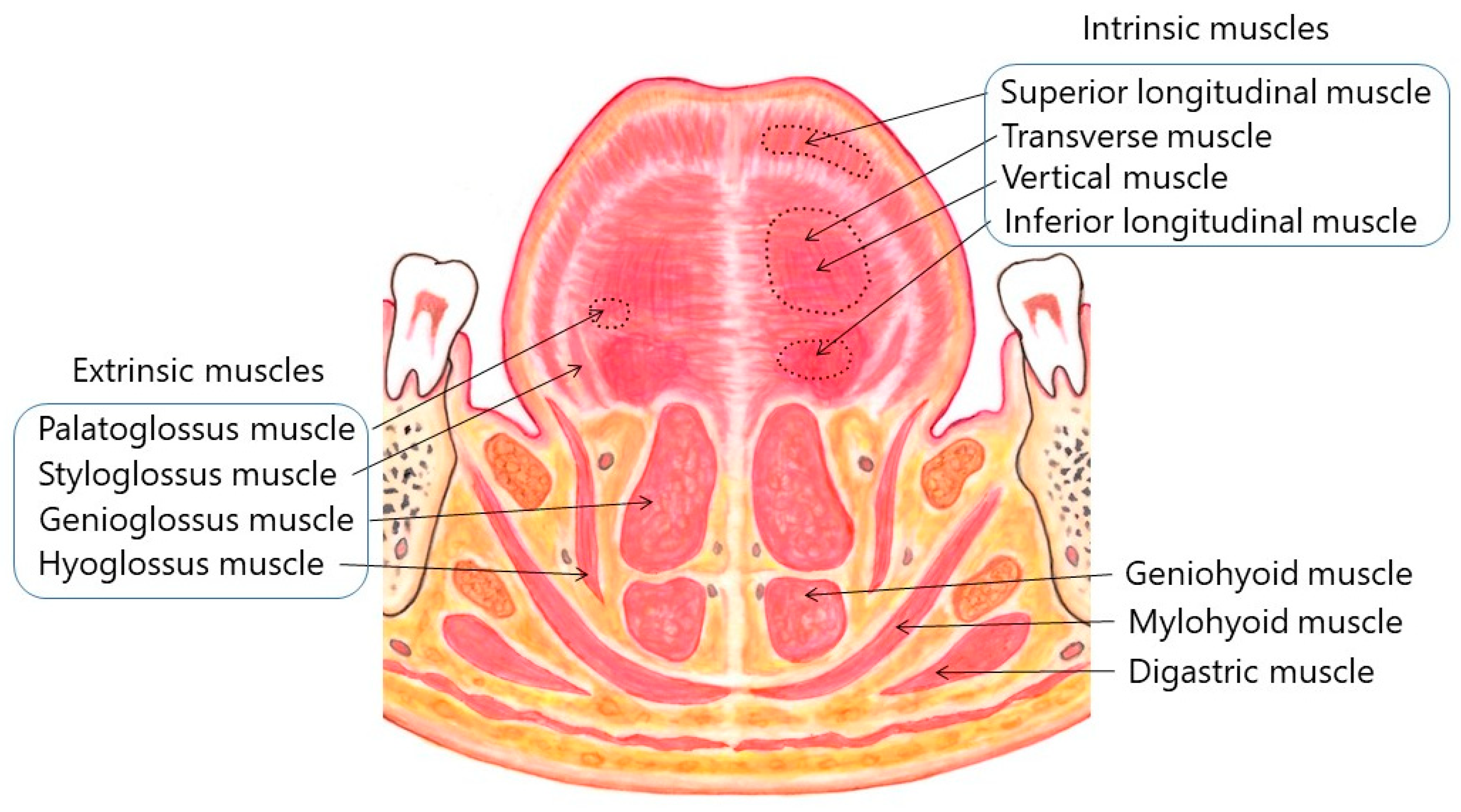
The tongue is a muscular structure located on the floor of the oral cavity. It is the primary taste organ and plays a key role in the initial phases of swallowing. In this article, we shall look at the anatomy of the tongue - its structure, innervation and clinical correlations. Intrinsic Muscles
Anatomical structure tongue taste buds on Vector Image
The tongue is a mobile, muscular organ that lies within the mouth and partly extends into the upper throat. The tongue's anatomy is complex; it involves interlacing muscles, nerves, and a blood supply.
Tongue Anatomy QA
1/2 Synonyms: none The tongue is a muscular organ situated in the oral cavity, and an accessory digestive organ. Its main functions include sensation of taste, mastication (chewing), deglutition (swallowing), speech, and clearing the oral cavity. The rich motor and sensory innervation of the tongue is carried by four cranial nerves

The Mouth, Pharynx, and Esophagus Anatomy and Physiology II
Tongue (Fig. 9.5) is made up of three elements; epithelium, muscles and glands. The epithelium is stratified and non-cornified. Two types of special structures are seen on it; the papillae (Fig. 9.6) and the taste buds. The taste buds (Fig. 9.7) are the sense organs of taste. These buds are lined by stratified squamous epithelium and are flask.
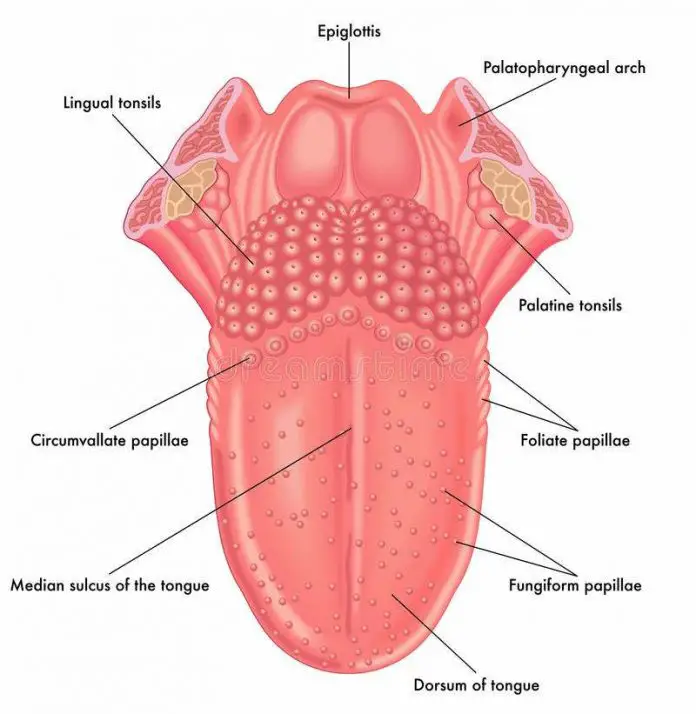
Diagram of tongue
Many parts make up your mouth anatomy. These parts work together harmoniously to help with chewing, speaking and breathing. The outside of your mouth creates a boundary that holds food in place and helps you form sounds and words. It includes your cheeks and lips. The inside of your mouth contains your: Teeth. Gums.

Print of Diagram illustrating the anatomy of the tongue, front view Anatomy of the tongue
Read about the human tongue and view a tongue diagram. Learn about the parts of the tongue, which includes taste buds, and learn about the tongue's function. Updated: 09/13/2022 What is.

Anatomy of Tongue Biology Ease
Anatomy Anterior two thirds Posterior third Muscles Histology The lingual papillae Taste buds Blood supply and lymphatic drainage Arteries Veins Lymphatic drainage Innervation Motor innervation
Hypogeusia definition, causes, symptoms, diagnosis, treatment & prognosis
Tongue | Anatomy, Parts, Pictures, Diagram of Human Tongue Posted by Dr. Chris The human tongue is a muscular organ that is covered by a thin mucous membrane. It lies partly in the mouth cavity and partly in the oropharynx. It is highly mobile and can be shifted into a number of different positions and also assume various shapes.